


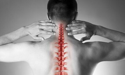
Cervical osteochondrosis or spondylosis develops as a result of changes in the shape and structure of the vertebrae.Even though the cervical region is quite short compared to the entire length of the spine, it is perhaps the most important part of the spinal column.Each of the adjacent vertebrae forms intervertebral foramina through which nerve roots emerge and are directed to all muscles and organs of the upper half of the body.The vital blood vessels that supply the brain pass through other openings - in the lateral processes of these vertebrae.
Causes of osteochondrosis of the cervical spine
The causes of osteochondrosis are as follows:
- injuries,
- "sitting" work on the monitor below eye level,
- physical work related to carrying heavy loads,
- drive a car for a long time,
- work "on the phone" without using remote devices (in this case, the operator presses the handset to his ear with his shoulder)
- constitutional characteristics (torticollis, congenital changes in the cervical vertebrae, short neck)
Development of pathological changes in the vertebrae
In the case of osteochondrosis, small sharp points begin to form on the edges of the vertebral bodies, which can injure nearby structures.Most often, this occurs due to excessive loading of the cervical spine, and is not only the result of "aging" of the intervertebral joints (remember that osteochondrosis used to be considered a degenerative, natural "age-related" disease, like osteoarthritis).As the disease progresses, the vertebral endplates become denser and the height of the intervertebral discs decreases.These discs usually act as shock absorbers between the vertebrae and, among other things, prevent damage to the spinal roots.In the case of progressive osteochondrosis, the nucleus pulposus of the intervertebral disc protrudes (hernia), which during the course of the disease is under increasing pressure, while the "holding" ligaments on all sides weaken.This hernia can also compress spinal structures and cause neurological manifestations of the disease.
What are the symptoms of cervical osteochondrosis?
Osteochondrosis of the cervical spine with pain syndrome
Any pain in the neck area suspects the pathology of the cervical spine.According to the increasing intensity of the pain syndrome, they can be divided into 4 stages, first the patient feels numbness, tingling, "tension" in the area of a certain muscle group, in the fourth stage - the most severe - the pain is so intense that it leads to the patient's immobility and reduced performance.
In addition to the pain in the neck and occipital region, the patient notices "referred" (radiating) pain in the upper limb and in the lateral areas of the chest below the scapula.
Osteochondrosis of the cervical spine with radicular syndrome
The involvement of the nerve roots in the process is indicated if pain, numbness and tingling spread to the lower jaw, upper back, forearm and fingers.At the same time, the patient notes that he "seemed to be resting" his hand and slept uncomfortably.There is morning stiffness in the joints of the fingers, which lasts no more than 10-15 minutes.With the development of radicular syndromes, a decrease in the muscle strength of the upper limbs can be observed during the examination.
Osteochondrosis of the cervical spine with "vertebral artery syndrome"
Involvement of blood vessels in the process (compression by a herniated disc or osteophytes) can be indicated if the patient complains of frequent headaches, especially when standing for a long time, when throwing the head back (for example, during breaststroke), tinnitus and dizziness.This clinical situation can be easily identified by ultrasound ("Doppler mapping mode").Ultrasound shows the tortuosity of the vertebral arteries and the narrowing of their lumen.In this case, we can talk about surgery, since a pronounced change in the blood flow of the vertebral arteries is a risk factor for stroke.
Osteochondrosis of the cervical spine with "heart (heart) syndrome"
This syndrome prompts the patient to consult a cardiologist first, since the main complaints are pain in the left side of the chest, under the shoulder blade, which weakens or intensifies when doing physical activity or changing body position.After heart attack and other heart diseases have been ruled out, the patient is admitted under the supervision and treatment of a neurologist and orthopedist.
Diagnostics
Four methods are used to clarify the diagnosis: radiography, ultrasound, computed tomography and magnetic resonance imaging.
The most accessible method is still radiography of the cervical spine;the most informative is the radiograph in the lateral projection ("side view").This method makes it possible to determine the presence of injuries and gross structural changes of the vertebrae with a first approximation.
An ultrasound examination (ultrasound) is performed to clarify the condition of the vertebral arteries.With this method, it can be determined whether the blood flow is impaired and, if so, to what extent and what obstacles have arisen and where they are located.
Computed tomography (CT).It allows for a more accurate assessment of the state of the bone structures, the level of bone tissue density, and enables the viewing of smaller osteophytes (bone growths) than is possible with radiography.
Magnetic resonance imaging (MRI).This type of examination is essential if there is a suspicion of the existence of a hernia, the exact location of the spinal cord damage and the extent of the damage.This examination is necessary when the question of operative (surgical) treatment of diseases of the cervical spine arises.
Treatment of cervical osteochondrosis
Drug treatment
The standard set of drugs for the treatment of cervical osteochondrosis reflects the goals of treatment: to relieve pain by eliminating painful muscle spasms and inflammation of the nerve roots, while increasing the mobility of the spine.In order to achieve these goals, pain relievers, NSAIDs - non-steroidal anti-inflammatory drugs, muscle relaxants - are used.It should be remembered that self-medication with drugs from these groups can be dangerous, as there is a possibility of misinterpreting the symptoms, as well as underestimating the side effects of these drugs.Topical (skin) NSAIDs are widely used in the form of gels, and when the pain is gone, the same drugs can be used in the form of ointments.
Slow-acting systemic drugs are used for deeper, "basic" treatment of osteochondrosis.These substances restore the cartilage structures of the vertebrae and prevent their further damage.Treatment courses are long, the effect lasts for months.
Cervical osteochondrosis shows significant differences from the pathology of other parts of the spine.In this case, the neck pain may not be caused by signals from the affected spinal nerves, but by painful, chronic muscle tension - all of this is called muscle tone syndrome.This is a completely "benign" condition that responds well to treatment with the same set of drugs: non-steroidal anti-inflammatory drugs, muscle relaxants, intramuscular "blockades" with steroids.Usually, the doctor notices a sharp pain when palpating the so-called "trigger" points along the entire cervical spine, as well as in the area of the muscles of the upper shoulder girdle.More often, this pathology occurs in women, most of whom are under 40 years of age.Despite the severe pain syndrome, the neurovascular structures remain intact and the blood flow in the head area is not affected.
Manual therapy
This treatment method can be effective in the case of neck pain that has recently occurred (often as a result of a minor injury or subluxation), which is not accompanied by dizziness or other changes in the nervous system and circulatory system.Manual therapy is allowed only after a thorough examination;in addition, the doctor performing the procedure must have sufficient experience in the field of traumatology and orthopedics.The use of manual therapy for "old" forms of the disease is dangerous!
There are two known methods of this type of intervention:
- manipulation (sharp short impacts with significant force, aimed at eliminating subluxations, the well-known "bone snaps");
- mobilization (the method is based on smooth stretching of the neck, after warming up and relaxing the muscular ligaments of the neck).
A combined method is also used, which is based on the combination of two main ones.It is important to note that, in addition to these contraindications, manual therapy is prohibited for all diseases associated with increased blood pressure, thyroid gland and any pathology of the ear, nose and throat organs.
Treatment of cervical osteochondrosis at home
Therapeutic exercises in cervical osteochondrosis
The first and main rule for beginners in physical therapy is not to do exercises while dealing with painful sensations.It goes without saying that you should not start in the "acute" period, when the pain has just appeared.Another important recommendation is to avoid sudden and circular movements of the cervical spine.
Each treatment should begin with a short, light self-massage of the neck muscles.
This is followed by a “warm-up” warm-up:
- The arms are lowered along the body, the shoulders are horizontal, the back is straight (you can check your posture by slightly pressing your heels, shoulder blades and buttocks against the wall).We walk in place for 1 minute on the whole leg, 1 minute on the toes, 1 minute on the heels.
- The initial situation is the same.We clench our hands into fists, raise and lower our shoulders, and keep our arms straight.The movements are slow, we do 20 repetitions, the last rise is 5 seconds longer.We make sure that the neck muscles are not strained.
- The initial situation is the same.We tilt our heads one by one to the right and then to the left.The movements are smooth, one tilt for 8 counts, at the extreme point of the tilt - hold for 8 seconds.
- The starting position is the same, or sitting on a hard chair.The head can be smoothly tilted forward, at the extreme point - hold for 8 seconds
- The starting position is the same, or sitting on a hard chair.Slowly tilt your head forward until your chin touches your chest, then slowly turn your head to the right (4 counts) and left (4 counts).Avoid overexerting your muscles.
- The starting position is the same, or sitting on a hard chair.We raise our shoulders for 4 counts, then lower them smoothly for 4 counts.10 repetitions.
- The starting position is the same, or sitting on a hard chair.We raise our shoulders, but now we make circular movements from front to back, to the count of 8.10 repetitions.
- We straighten our backs and check our posture.For a count of 4, bring your shoulder blades together behind your back, try to connect them, linger for 8 seconds at the end point, and then return to the starting position.
Pillows
As already mentioned, hypertonia of the neck muscles is the first and often the main reason for the development of cervical osteochondrosis.Reasonable selection of pillows and mattresses and ensuring a calm and comfortable sleep are no less important than gymnastics, physical therapy and medicines.
When choosing a mattress, pay attention to the composition of the filler (products that are at least half made of coconut shavings, i.e. have sufficient stiffness).Soft spring mattresses do not ensure proper alignment of the spine.The most optimal sleeping position is on your side, with one or both knees pulled up towards your stomach.The pillow should be placed so that it fills the entire space between the shoulder, the ear and the mattress, while the parietal part of the head (crown) is in a horizontal line with the spine.Pillows that are too high and too low, as well as soft, should be avoided.The ideal solution is an ergonomically shaped product, i.e. in this case with a small pressure cylinder on one side.
General recommendations
Pay attention to your posture.When walking or standing, the correct position is when the chest is extended and the stomach is pulled in.
Avoid sitting for long periods of time.A simple rule is known to prevent cervical osteochondrosis: after every 60 minutes of work, a 10-15 minute walk or warm-up is required.
The work chair must have a high headrest or backrest.
While sitting, your feet should lie on the floor and your neck should not be strained.For this purpose, use special orthopedic devices: a support under the neck, if driving in a car, a pillow under the back.
Avoid heavy lifting.If necessary, kneel down, hold a heavy object to your torso, then stand up smoothly, using the strength of your leg muscles, but not the "pull" of your back.
Do not bend with straight legs.Use tripods or work surfaces to bring your subject closer instead of leaning your face toward the subject.Try doing your homework sitting on a chair or an exercise ball.
If you have to use a mop, broom or rake, do not strain your arms, back, neck or lean to one side.
Avoid breaststroke.