


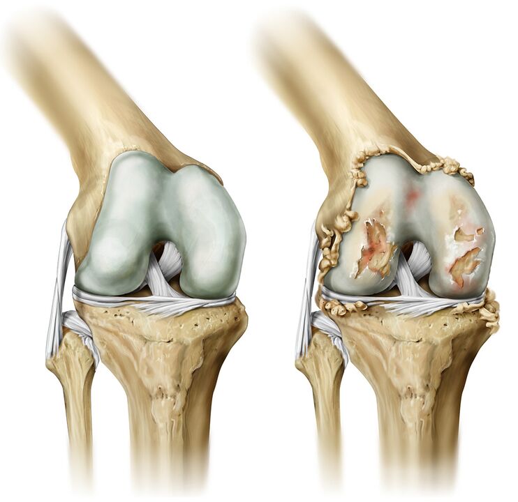
Arthrosis of the joints is a chronic disease characterized by the development of degenerative lesions of the articular cartilage, resulting in deformation of the bone tissue. The joints of the big toes, hips, and knees are most commonly affected.
Symptoms of the disease
- The first clinical symptom of arthrosis is pain in the affected joint during excessive physical exertion. Painful feelings may occur during movement. As the disease progresses, joint pain also disturbs the person at rest and causes insomnia.
- Cracking of the joints. The destruction of the cartilage layer results in friction of the bones, with clicks and cracks when moving in the joint. As the disease progresses, the crisis intensifies.
- Reduced mobility. If the joint is damaged, movements are restricted, in the case of severe arthrosis, the patient has limb stiffness in the morning.
- Joint deformity. Without proper and timely treatment, the joint will deform and its appearance will change.
- As the inflammatory process worsens, the patient decreases the sensitivity of the toes and numbness of the fingertips.
Causes of the disease
The main cause of arthrosis is an increase in the cartilage layer between the joint and the bone. The contributing factors are:
- Intense physical activity;
- Joint microtrauma;
- Common fractures
- Wear tight shoes or high heels
- Congenital tendency.
Diagnostics
The main method of diagnosing arthrosis is a carefully collected patient history (professional history).
Diagnosis is made based on examination of the patient and further examinations, including x-rays of the joints, arthroscopy, ultrasound, MRI, and computed tomography.
- Ultrasound. This research method is reliable and harmless. Because ultrasound diagnosis refers to non-invasive methods, there are no contraindications to this study. Ultrasound can be used to diagnose thinning of cartilage tissue, degenerative changes in the joint meniscus, thickening of the joint membrane, and the presence of fluid in the joint cavity. This study allows for accurate selection of arthrosis treatment.
- MRI and computed tomography. Computed tomography and MRI can be used to assess the condition of the joint: cartilage thickness, erosions, or cysts in the bone tissue to determine the amount of intraarticular fluid.
- Arthroscopy. This test is done more often to determine the cause of arthrosis.
Complications
In the absence of timely medical care, arthrosis progresses and threatens with complications such as:
- Inflammation of the tissues around the joint;
- Restriction of mobility of affected joints;
- Degenerative changes in the hip joint;
- Changing the shape of joints.
Treatment of the disease
Treatment is prescribed to the patient depending on the degree of joint damage. Therapy for arthrosis begins with pain relief.
In parallel with painkillers, the patient is prescribed anti-inflammatory drugs. In addition to medication, the patient undergoes a course of physiotherapy.
Massaging the affected limbs after the acute form of the inflammatory process has subsided can reduce pain, normalize joint mobility, and relieve muscle cramps.
Physiotherapy exercises are prescribed to relieve muscle stiffness, warm up, and strengthen the patient’s general condition. Exercise helps maintain proper posture and smooth walking.
Sanatorium treatment is indicated during periods of stable remission. Mud baths, applications and other procedures help restore joint motor function and relieve pain.
If conservative treatment methods do not produce the expected effect, the patient will be prescribed surgical joint replacement. Endoprostheses are made of a material that is not rejected by the human body. They allow the physiological functions of the affected joint to be fully restored.
Individual treatments: radiofrequency ablation and disruption of method integrity by disruption of the integrity of the pain-causing nerve.
Risk group
People at risk include:
- Overweight;
- Varicose veins;
- Athletes;
- Pianists;
- Programmers.
Prophylaxis
The prevention of arthrosis is as follows:
- Good nutrition;
- Prevention of injuries and fractures;
- Limiting the load on the joints with hereditary predisposition;
- Weight control;
- Wear suitable shoes.
Diet and lifestyle
Diet should be adjusted as hereditary predisposition to arthrosis develops and the disease progresses. It is recommended to include sea fish (sardines, salmon, tuna), fresh vegetables and fruits, cereals in the diet. Limit cakes, fatty meats, chocolate and alcohol.
We recommend that you spend more time in the fresh air and do not expose your joints to increased physical activity.